Anatomical Relations
👩⚕️ Work through these high yield Q&As. Try to learn everything here!
Which part of the small bowel does an annular pancreas affect?
2nd part of the duodenum
🔖 Annular pancreas is a rare congenital anomaly where a congenital ring of pancreatic tissue encircles the 2nd part of the duodenum, potentially leading to duodenal obstruction. It occurs due to abnormal migration of the ventral pancreatic bud during embryological development.
Which renal vein passes anterior to the abdominal aorta before draining into the IVC?
Left renal vein
🔖 The left renal vein is long, crossing anterior to the aorta to drain into the IVC. Nutcracker syndrome is where the left renal vein is compressed between the abdominal aorta and the superior mesenteric artery causing venous hypertension, left flank/pelvic pain, haematuria, proteinuria, and varicocele.
👩⚕️ These are MCQs. Try to answer them then hover over the options to check your answer and read the explanations.
Which structure is not in direct posterior relation to the 3rd part of the duodenum?
- ((Right kidney::☑️ Sits higher, T12 to L3, and more laterally; posterior to the 2nd part of the duodenum))
- ((Right ureter::Right ureter is posterior to the 3rd part of the duodenum))
- ((Aorta::Aorta is posterior to the 3rd part of the duodenum))
- ((IVC::IVC is posterior to the 3rd part of the duodenum))
🥪 The 3rd part of the duodenum is retroperitoneal and runs horizontally at about L3, from right → left, and is famously sandwiched:
Anterior to the 3rd part of the duodenum:
- Superior mesenteric artery (SMA)
- Superior mesenteric vein
- Root of mesentery
Posterior to the 3rd part of the duodenum:
- Abdominal aorta
- Inferior vena cava
- Right ureter
- Right psoas major
- Sympathetic chains
What helps the surgeon identify the uncinate process of the pancreas?
- ((Lies anterior to inferior mesenteric artery::No meaningful direct relationship between the IMA and the uncinate process))
- ((Lies anterior of superior mesenteric artery::The uncinate process hooks posterior to the SMA/SMV at about L1–L2))
- ((Lies posterior to superior mesenteric artery::☑️ SMA and SMV sit anterior to the uncinate process of the process))
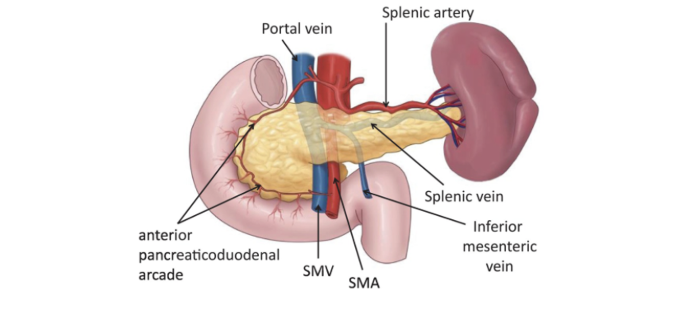
Which of the following lies anterior to the head of the pancreas?
- ((Stomach pylorus::☑️ Pylorus, transverse colon and transverse mesocolon are anterior to pancreas head))
- ((IVC::IVC, right renal vessels, and CBD run posterior to the pancreas head))
- ((Gastroduodenal artery::Gastroduodenal artery runs superior to the upper border of the pancreas head))
Which structure lies immediately inferior to the left adrenal gland?
- ((Spleen::The stomach, body of pancreas, spleen and upper pole of left kidney lie anterior to the left adrenal gland))
- ((Stomach::The stomach, body of pancreas, spleen and upper pole of left kidney lie anterior to the left adrenal gland))
- ((Body of pancreas::☑️ The body of the pancreas is primarily anterior but also slightly inferior in vertical level, to the left adrenal gland))
- ((Left renal vein::The left renal vein runs horizontally from the kidney to the IVC, crossing anterior to the aorta, and inferior to the left adrenal gland))
Which structure lies immediately anterior to the right adrenal gland?
- ((Stomach::The stomach, body of pancreas, spleen and upper pole of left kidney lie anterior to the left adrenal gland))
- ((Spleen::The stomach, body of pancreas, spleen and upper pole of left kidney lie anterior to the left adrenal gland))
- ((Pancreas::The stomach, body of pancreas, spleen and upper pole of left kidney lie anterior to the left adrenal gland))
- ((Inferior vena cava::☑️ The inferior vena cava, right lobe of the liver and upper pole of right kidney lie anterior to the right adrenal gland))
🫷 These lie anterior to the right adrenal gland
- inferior vena cava
- right lobe of the liver
- upper pole of right kidney
🫸 These lie anterior to the left adrenal gland
- stomach
- body of pancreas
- spleen
- upper pole of left kidney
A 53-year-old man with a chronically infected right kidney is due to undergo a nephrectomy. Which of the following structures would be encountered first during a posterior approach to the hilum of the right kidney?
- ((Right renal artery:: Encountered after the ureter, in the middle))
- ((Ureter::☑️ Structures at the right renal hilum from anterior to posterior: Renal vein → Renal artery → Ureter))
- ((Right renal vein:: Lies anterior to the renal artery and ureter, thus encountered last))
- ((Right testicular vein::Not encountered at the hilum))
Which describes the correct anatomical relationship of the internal iliac artery, internal iliac vein and ureter?
- ((Internal iliac vein is anterior to the internal iliac artery::The internal iliac is anterior to the internal iliac vein))
- ((Ureter is anterior to the internal iliac artery::☑️ The ureter is anterior to the internal iliac artery))
- ((Internal iliac artery is posterior to the the internal iliac vein::The internal iliac artery is anterior to the internal iliac vein))
⭐ From anterior to posterior on the pelvic sidewall: Ureter → internal iliac artery → internal iliac vein
Which describes the correct anatomical relationship of the internal iliac artery, internal iliac vein and ureter?
- ((Internal iliac vein is anterior to the internal iliac artery::The internal iliac is anterior to the internal iliac vein))
- ((Internal iliac artery is anterior to the internal iliac vein::☑️ The internal iliac artery is anterior to the internal iliac vein))
- ((Internal iliac artery is posterior to the the internal iliac vein::The internal iliac artery is anterior to the internal iliac vein)
⭐ From anterior to posterior on the pelvic sidewall: Ureter → internal iliac artery → internal iliac vein
Which of the following statements correctly describes the anatomical relations of the ureter?
- ((The ureter is crossed posteriorly by the gonadal veins::No, water goes under the bridge))
- ((The ureter passes posterior to the bifurcation of the common iliac arteries::Ureter crosses anterior to/over the internal iliac artery near the pelvic bridge))
- ((The ureter is crossed anteriorly by the gonadal veins::☑️ The ureter passes under the gonadal vessels in the abdomen (water under the bridge)))
- ((The ureter lies lateral to the psoas major muscle::The ureter lies on the anterior surface of psoas major))
🌉 Water runs under the bridge: The ureters pass under the gonadal vessels in the abdomen and the ureters pass under the uterine or vas deferens vessels in the pelvis.
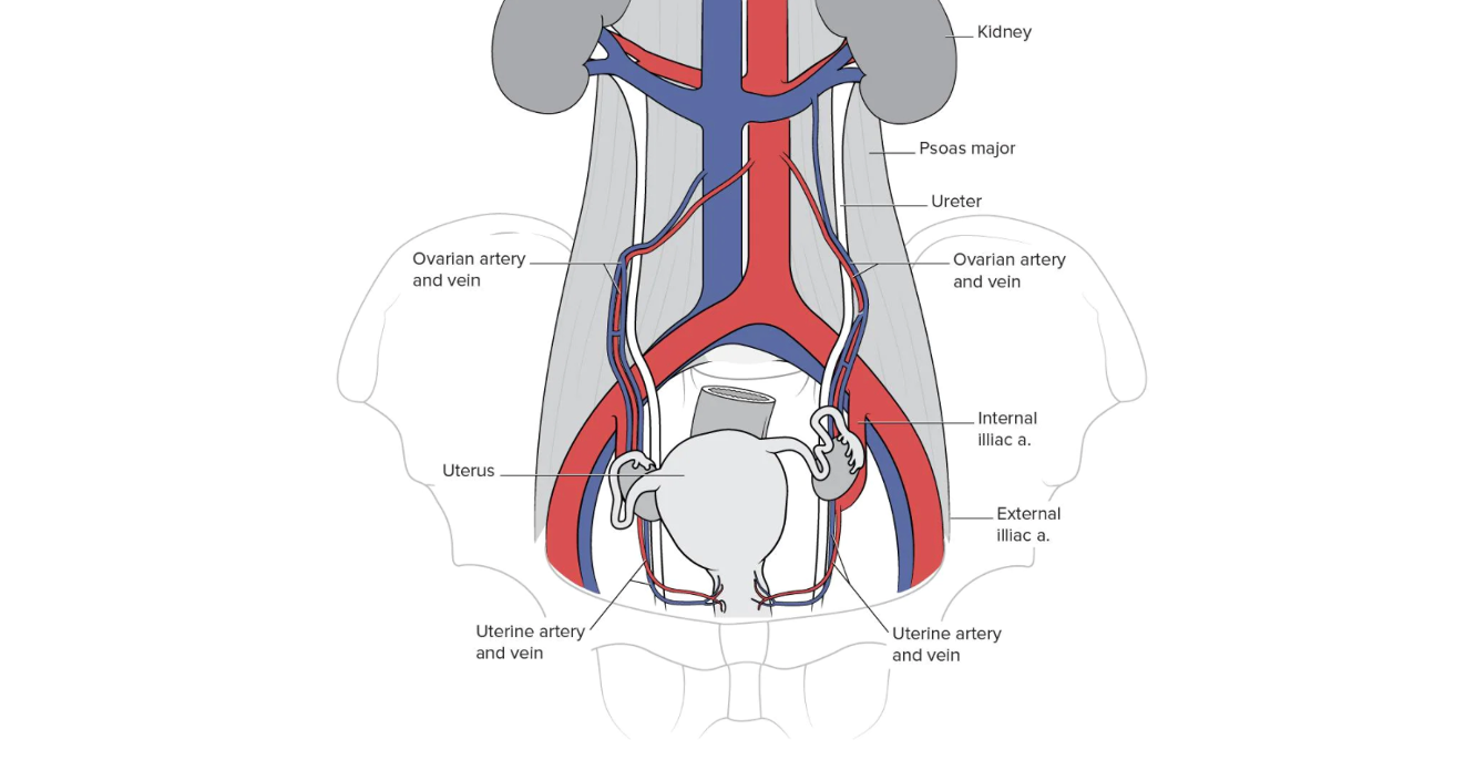
Which of the following statements correctly describes the anatomical relations of the ureter?
- ((The ureter is crossed posteriorly by the gonadal veins::No, water goes under the bridge))
- ((The ureter passes posterior to the bifurcation of the common iliac arteries::Ureter crosses anterior to/over the internal iliac artery near the pelvic bridge))
- ((The common iliac arteries divide posterior to the ureter::☑️ The left and right ureters lie anterior to the terminal bifurcation of the left and right common iliac arteries))
- ((The ureter lies lateral to the psoas major muscle::The ureter lies on the anterior surface of psoas major))
🎋 The terminal bifurcation of the left and right common iliac arteries are crossed anteriorly by the left and right ureters.
At which part of the vertebra does the ureter insertion into the bladder occur in relation to vertebral anatomy?
- ((At the level of L1-L2 disc::The ureter typically begins at the L2 level))
- ((Anterior to L3 vertebral body::As it descends from the renal pelvis, the abdominal portion of the ureter runs retroperitoneally along the anterior surface of the psoas major muscle. The psoas lies in front of the transverse processes and bodies of the lumbar vertebrae.
- ((At the level of the iliac crest:: The highest point of the iliac crest corresponds to L4 spinous process (the classic Tuffier’s line).The ureter crosses into the pelvis just below the level of the iliac crest.
- ((Anterior to the transverse process of L5::The ureter does pass anterior to the transverse processes of L5 as it descends on the psoas major toward the pelvic brim))
- ((Posterior to the sacroiliac joint::As the ureter approaches the pelvic brim, it leaves the psoas major muscle and moves medially toward the sacroiliac joint. The ureters pass directly in front of the sacroiliac joint as they transition from the abdomen to the pelvic cavity. At this level, (≈ L5/S1), the ureter crosses the bifurcation of the common iliac artery. The artery is posterior to the ureter, and the SI joint is posterior to the artery.
- ((Anterior to the sacrum at S2::☑️ The ureters enter the urinary bladder at the posterolateral angles of the trigone, at S2 level))
🛣️ Anatomical course of the ureters
- The ureters arise from the ureteropelvic junction at the renal pelvis at approximately the level of the L1–L2 vertebrae
- They descend retroperitoneally on the anterior surface of the psoas major muscle, passing in front of the lumbar transverse processes and lying anterior to the sympathetic chain and genitofemoral nerve.
- As they descend, they pass anterior to the transverse process of L5, then reach the pelvic brim
- At the pelvic brim, around the level of L5–S1 and opposite the sacro-iliac joint, each ureter crosses anterior to the bifurcation of the common iliac artery (or occasionally the external iliac artery) to enter the pelvis.
- Within the pelvic cavity the ureters descend along the lateral pelvic walls, anterior to branches of the internal iliac artery and the obturator neurovascular bundle, roughly from S1 to S3.
- At the level of the ischial spines (around S3) they turn anteromedially and run transversely toward the base of the bladder.
- Approximately 2 cm above the ischial spine, the ureter passes beneath the uterine artery in females or beneath the vas deferens in males.
- Finally, the ureters enter the posterolateral wall of the bladder obliquely at about the level of the S2 vertebra, forming the superior corners of the trigone; this oblique intramural course acts as a functional one-way valve to prevent vesicoureteric reflux.
Which of the following is NOT a posterior relation of the kidney?
- ((Iliacus::☑️ The iliacus muscle lies lower in the iliac fossa, in the pelvis))
- ((Psoas major::Psoas major runs vertically along the lumbar vertebral bodies and forms part of the posterior abdominal wall directly behind the medial aspect of each kidney))
- ((Quadratus lumborum::Quadratus lumborum lies lateral to psoas major and posterior to the kidney, forming a major component of the posterior abdominal wall))
- ((Transversus abdominis::Transversus abdominis posterior fibers and aponeurosis contribute to the posterior abdominal wall and lie behind the kidney))
- ((Diaphragm::The upper poles of the kidneys lie directly beneath the diaphragm; the diaphragmatic pleura and costodiaphragmatic recess are clinically relevant during renal surgery)