Embryology
Germ Layers & Vertebral Development
- Vertebrae develop from sclerotome, a derivative of the paraxial mesoderm (somites).
- Failure of complete formation or segmentation of the sclerotome leads to hemivertebra, an important cause of congenital scoliosis.
- Ectoderm forms skin and nervous system structures
- Endoderm forms intestinal epithelial linings
- Notochord does not form vertebrae directly; it induces vertebral column development
- Neural tube develops into brain and spinal cord
Pharyngeal arches
Pharyngeal arches are transient bulges that appear on the lateral aspect of the embryonic head region between day 27 to 29. Between weeks 5 to 8, the mesenchyme differentiates into skeletal, muscular, and vascular derivatives.
Pharyngeal pouches are endodermal evaginations between the arches, giving rise to internal structures.
- First pouch gives rise to the tympanic cavity and auditory tube
- Second pouch gives rise to the palatine tonsils lining and the tonsillar fossa
- Third pouch gives rise to the inferior parathyroids and thymus
- Fourth pouch gives rise to the superior parathyroids and ultimobranchial body
Pharyngeal clefts normally regress except the first cleft which forms the external auditory meatus.
Persistence or malformation of pharyngeal arch structures yield congenital syndromes such as Treacher‑Collins (1st arch neural‑crest dysfunction), Di George (3rd & 4th pouch aplasia), and branchial cleft cysts/fistulae.
During the 6th week of gestation, the second pharyngeal arch overgrows the third and fourth arches, creating a temporary ectoderm-lined depression known as the cervical sinus. This sinus is meant to fuse and be reabsorbed. A branchial cleft cyst is a congenital fluid-filled sac that forms when the cervical sinus from this second arch fails to fully obliterate.
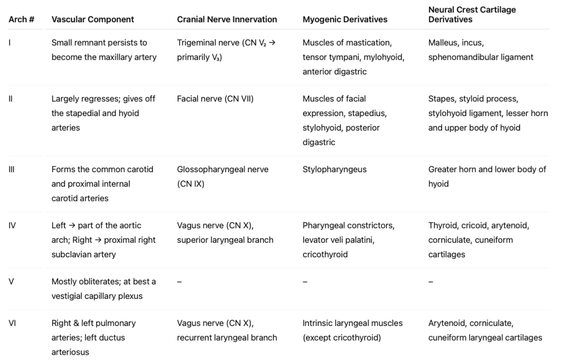
Derivations of the pharyngeal arches
👩⚕️You must memorise this table. It's not as hard as it may look at first glance! Perhaps create mini flashcards to help?
👩⚕️ Now work through these Q&As.
Hemivertebra develops from which embryological structure?
Mesoderm develops into a range of tissues including muscles, bone, cartilage, blood, connective tissues, the circulatory system, the urogenital system and dermis of the skin and the lining of the body cavity
The muscles of mastication develop from which pharyngeal arch?
The muscles of mastication originate from the first pharyngeal arch and are all innervated by the trigeminal, CN V3. These include the temporalis, masseter, medial and lateral pterygoids.
A patient presents with unilateral facial weakness. From which pharyngeal arch does the affected nerve originate?
The muscles of facial expression originate from the second pharyngeal arch and are all innervated by the facial nerve, CN VII.
🔖 CN 5 - 7 - 9 innervate the 1st - 2nd - 3rd arches; The SLN and RLN of CN 10 innervate the 4th and 6th arches
A 21-year-old man undergoes an uncomplicated tonsillectomy for recurrent tonsillitis. Postoperatively, he complains of otalgia despite no ear pathology. The nerve responsible for this referred pain originates from which pharyngeal arch?
The glossopharyngeal CN IX, from the 3rd pharyngeal arch.
🔖 The glossopharyngeal CN IX innervates the 3rd pharyngeal arch. It supplies the tonsils, posterior third of the tongue, and the tympanic plexus. Referred otalgia after tonsillectomy occurs due to shared sensory innervation between the tonsillar fossa and the middle ear via the glossopharyngeal CN IX, from the 3rd pharyngeal arch.
What is the embryological arch of origin of the ductus arteriosus?
The left 6th pharyngeal arch artery persists to form the ductus arteriosus, connecting the pulmonary artery to the aortic arch in the fetal circulation.
🔖 The left 6th pharyngeal arch artery persists to form the ductus arteriosus, connecting the pulmonary artery to the aortic arch in the fetal circulation. The 6th pharyngeal arch also gives rise to the left and right pulmonary arteries.
Memorise these vascular derivations of the pharyngeal arches:
- First arch ➡️ Maxillary arteries
- Second arch ➡️ Stapedial artery
- Third arch ➡️ Common carotid and internal carotid arteries
- Fourth arch ➡️ Aortic arch, right subclavian
- Sixth arch ➡️ Left and right pulmonary arteries and ductus arteriosus
What is the embryological arch of origin of the left and right pulmonary artery?
6th pharyngeal arch
🔖 The 6th pharyngeal arch gives rise to the left and right pulmonary arteries, and the ductus arteriosus.
The inferior parathyroid gland is derived from which structure?
The third pouch gives rise to the inferior parathyroids and thymus.
🔖 The 3rd pouch gives rise to the inferior parathyroids and thymus. The 4th pouch gives the superior parathyroids and ultimobranchial body.
A cervical sinus forms from which branchial arch?
A cervical sinus forms from the second arch.
A branchial cyst is a derivative of which structure?
The branchial cyst is a derivate of the second arch.
🔖 A branchial cleft cyst is a congenital fluid-filled sac that forms when the cervical sinus fails to fully obliterate. It is a remnant of this embryonic structure that persists after birth.
A pharyngeal pouch (Zenker’s diverticulum) arises through a weak area in the posterior pharyngeal wall. Between which two muscles does it occur?
Inferior pharyngeal constrictors: thyropharyngeus and cricopharyngeus
🔖 A pharyngeal pouch (Zenker’s diverticulum) arises through a weakness in the posterior pharyngeal wall between the thyropharyngeus and cricopharyngeus; both form the inferior pharyngeal constrictor muscle.
Where is a pharyngeal (Zenker’s) pouch located?
Through the inferior constrictors, between the thyropharyngeus and cricopharyngeus.
🔖 The Killian dehiscence is a triangular area of weakness formed by the oblique fibres (thyropharyngeus), and the transverse fibres (cricopharyngeus) of the inferior pharyngeal constrictor. Upper GI endoscopy is contraindicated for pharyngeal pouch and an UGI fluoroscopic swallowing study is done instead.
During fetal development, at what gestational age do the intestines return to the abdominal cavity after physiological herniation into the umbilical cord?
10 weeks
🔖 During normal embryonic development, the midgut herniates into the umbilical cord around week 6 due to rapid growth and limited intra-abdominal space. By week 10 to 11, the intestines retract back into the abdomen. Failure of this process results in omphalocele.
Which of the following is the axis of gut rotation during development?
Superior mesenteric artery.
🔖 The midgut herniates into the umbilical cord around week 6, then returns to the abdomen by week 10. While outside the abdominal cavity, the midgut rotates 270° counterclockwise around the superior mesenteric artery.
Failure of development of the caudal portion of the metanephros results in which condition?
Renal agenesis
🔖 The metanephros gives rise to the definitive kidneys and if the caudal portion of the metanephros fails to develop properly, it can result in renal agenesis, meaning one or both kidneys fail to form.
An atrioventricular septal defect results from a developmental defect in which embryological structure?
Endocardial cushion
🔖 Endocardial cushions are crucial for forming the lower atrial septum, upper ventricular septum, and the atrioventricular valves, and their failure to fuse properly leads to the characteristic defects seen in AVSD.
A child presents with epispadias and bladder exstrophy. What is the embryological origin of the bladder?
The urogenital sinus is an endodermal structure giving rise to bladder (except trigone which derives from mesonephric duct).
🔖 Bladder exstrophy and epispadias arise from the same underlying embryological defect: a failure of mesoderm to migrate into the midline of the lower abdominal wall so the infra-umbilical abdominal wall cannot close properly. At the same time, the genital tubercle develops in an abnormally dorsal position, which prevents the urethral plate and genital folds from fusing normally. As a result, the urethra fails to form on the ventral surface and instead opens dorsally, producing epispadias. The combination of these defects — poor mesodermal migration and dorsal displacement of the genital tubercle — also prevents the anterior wall of the bladder from forming, leaving the posterior bladder wall exposed on the abdominal surface. This produces bladder exstrophy.
Hypospadias results from a developmental defect involving which of the following structures?
Urogenital folds.
🔖 Hypospadias occurs due to failure of the urogenital folds to fuse properly.
Failure of fusion of the
- urogenital fold - leads to hypospadias
- urogenital sinus - leads to epispadias and bladder exstrophy