Planes, Levels & Surface Markings
Key anatomical landmarks and structures by vertebral level
👩⚕️ Take time to memorise this table
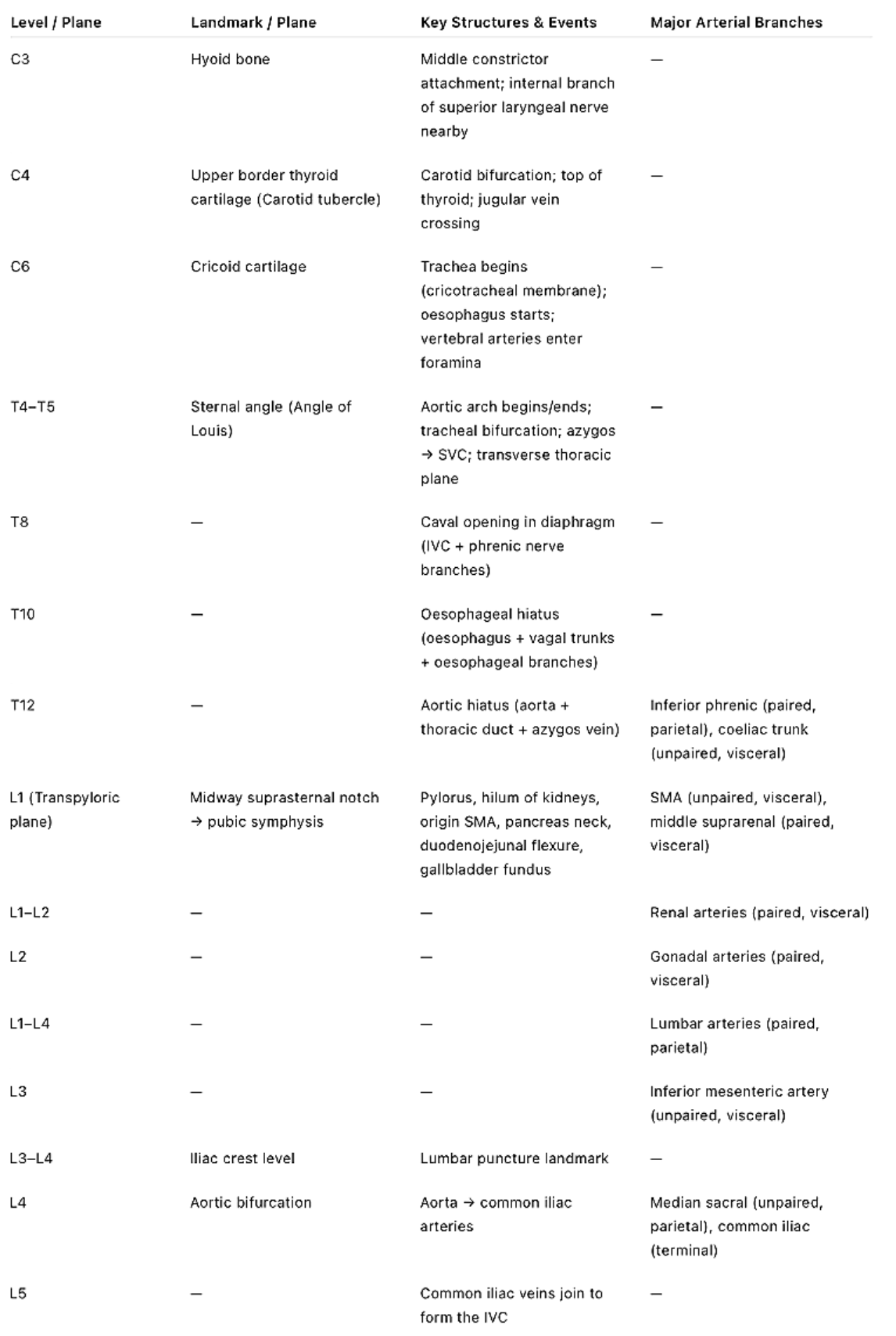
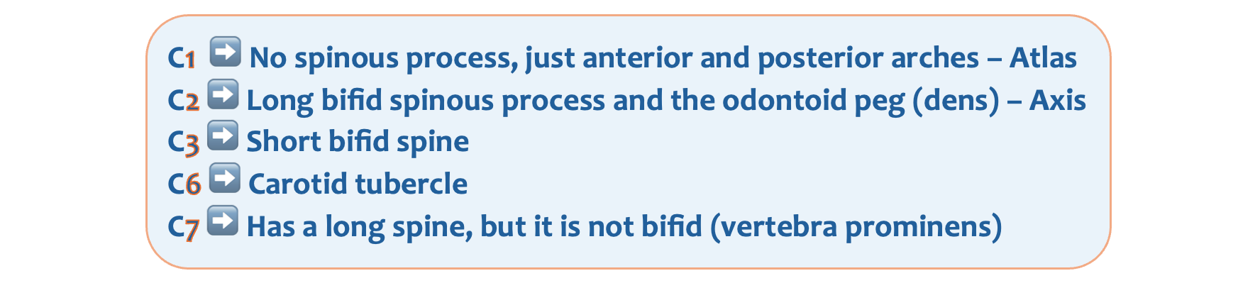
Cervical vertebrae
- C1 (atlas) lacks a spinous process; it only has anterior and posterior arches.
- C2 (axis) has a long spinous process and bears the odontoid process (dens).
- C3 features a short, bifid spinous process.
- C6, marked by its prominent carotid tubercle, is the level where the vertebral artery enters the transverse foramen, ascending through the cervical transverse foramina toward the skull
- At C6, the common carotid artery pulsation can be palpated, and the trachea terminates.
- The C7 vertebra, known as the vertebra prominens, has a long spinous process that is the most palpable vertebral landmark at the back of the neck.
The angle of Louis
The angle of Louis (sternal angle) at level T4/T5 is a key anatomical landmark. It corresponds to the following:
- Bifurcation of trachea (carina)
- NB The pulmonary trunk bifurcates below the trachea bifurcation at around the T5 vertebral level.
- Arch of aorta begins and ends
- Ligamentum arteriosum
- Azygous vein drains into SVC
- Thoracic duct crosses to left side
- Left recurrent laryngeal nerve hooks around arch (near ligamentum arteriosum)
- Marks the division between superior and inferior mediastinum
The azygos vein
The azygos vein begins at the level of L1/L2 from the right ascending lumbar and subcostal veins, entering the thorax through the aortic hiatus (T12). It ascends along the right side of the vertebral column, collecting blood from posterior intercostal and mediastinal veins. At about T4, the azygos vein arches over the right main bronchus and makes contact with the T5 vertebral body as it does so, and drains into the superior vena cava, providing an important collateral pathway between the SVC and IVC.
The three major openings of the diaphragm
- Caval hiatus at T8
- Oesophageal hiatus at T10
- Aortic hiatus at T12

- Structures passing through the caval hiatus at T8:
- IVC
- Branches of right phrenic nerve
- Structures passing through the oesophageal hiatus at T10:
- Oesophagus
- Anterior (mainly left vagus) and posterior (mainly right vagus) vagal trunk
- Oesophageal branches of left gastric vessels
- Lymphatics from lower oesophagus
- Structures passing through the aortic hiatus at T12:
- Aorta
- Thoracic duct
- Azygos vein
- Sometimes hemiazygos
Structures at the transpyloric plane at L1
- Pylorus of stomach (hence the name)
- Duodenojejunal flexure - should be located to the left at the transpyloric plane. Malposition of the DJ flexure to the right is a sign of malrotation.
- Fundus of gallbladder
- Hila of kidneys
- Origin of superior mesenteric artery SMA
- Portal vein formation behind neck of pancreas
- Pancreatic neck & body
- Cisterna chyli
- Tips of 9th costal cartilages
- Conus medullaris (end of spinal cord in adults)
👩⚕️ Now let's work through these high yield Q&As. Try to learn everything here!
What are the surface markings for the internal jugular vein?
Lobule of the ear to the sternoclavicular joint
What are the surface markings for the external jugular vein?
Angle of mandible to middle third of clavicle
What are the surface markings for femoral artery access?
Midpoint between the ASIS and pubic symphysis
What are the surface markings of McBurney’s point?
⅓ from ASIS to umbilicus
At what level does the common carotid artery bifurcate?
C4
This is also where the carotid sinus and body are located.
A child inhales a peanut. Where is it most likely to lodge?
Right lower lobe bronchus
The right main bronchus is wider, shorter, and more vertical than the left, so foreign bodies preferentially enter it, especially into the lower lobe bronchus.
Which cervical vertebra is characterised by a long bifid spinous process?
C2 (Axis) has a long bifid spinous process and the odontoid peg (dens).
The vertebral artery arises from the subclavian artery and ascends through the transverse foramina of the cervical vertebrae. It enters the foramen transversarium (transverse foramen) at which level?
C6
C6, marked by its prominent carotid tubercle, is the level where the vertebral artery enters the transverse foramen, ascending through the cervical transverse foramina toward the skull
At which vertebral level can the common carotid artery pulsation be felt, and the trachea ends?
C6
At C6, the common carotid artery pulsation can be palpated, and the trachea terminates.
Which cervical vertebra is most prominently felt on the back of the neck?
C7
Vertebra prominens, most palpable posteriorly
What is the level of the oesophageal opening in the diaphragm?
T10
Oesophagus has 10 letters!

Which structures pass through the oesophageal hiatus of the diaphragm?
- Oesophagus
- Vagus CN X
- Oesophageal branches
Which structures pass through the diaphragmatic opening at T12?
- Aorta
- Thoracic duct
- Azygos vein
- Hemiazygos
What is the level of the aortic hiatus of the diaphragm?
T12
Aortic hiatus has 12 letters so it occurs at T12.
Where is the duodenojejunal (DJ) flexure located under normal anatomical conditions?
On the left at the transpyloric plane
Left is normal, but right would indicate malrotation.
At which level is the neck of the pancreas?
The neck of the pancreas sits directly on the transpyloric plane at L1.
Structures at the transpyloric plane at L1
- Pylorus of stomach (hence the name)
- Duodenojejunal flexure
- Fundus of gallbladder
- Hila of kidneys
- Origin of superior mesenteric artery SMA
- Portal vein formation behind neck of pancreas
- Pancreatic neck & body
- Cisterna chyli
- Tips of 9th costal cartilages
- Conus medullaris (end of spinal cord in adults)
At what level does the aorta bifurcate?
L4
The aorta bifOURcates at LFOUR.
What is the best level for a lumbar puncture in a 2-year-old?
L4/L5
The spinal cord ends lower in infants (L3) than in adults (L1/L2).
A man presents with a DVT. CT confirms the presence of a thrombus in his common iliac vein extending into the inferior vena cava. The common iliac veins join to form the inferior vena cava at which vertebral level?
The left and right common iliac veins join to form the IVC at level L5.

At what level does the arch of aorta begins and end?
The angle of Louis (sternal angle) at T4/T5
At what level does the trachea bifurcate?
The angle of Louis (sternal angle) at T4/T5
👩⚕️ And here are a few MCQs to finish off.
A person sustains a stab wound just to the right of the angle of Louis, T4/T5. Which structure is not likely to be damaged?
- ((Right vagus::The right vagus lies in the mediastinum near the trachea and great vessels at this level, making it vulnerable to a penetrating injury at T4/T5.))
- ((Right pleura::The pleura extends anteriorly and laterally at this level, so a stab wound just right of the sternum can penetrate the pleural cavity.))
- ((Thoracic duct::Although mostly left-sided, the thoracic duct crosses to the right at T4–T5 before arching left again; therefore it can be injured by a wound at this level.))
- [[Brachiocephalic vein::The right brachiocephalic vein lies superiorly behind the sternoclavicular joint, above the T4/T5 plane, making damage at the angle of Louis unlikely.]]
The right brachiocephalic vein is higher up lying behind the sternoclavicular joint. It is formed by the fusion of the internal jugular and subclavian vein and courses obliquely down to join the left brachiocephalic to form the superior vena cava.
A structure located at the level of the T5 vertebra is:
- ((Left atrium::The left atrium lies posterior to the oesophagus at around T6 to T8))
- ((Tracheal bifurcation::Tracheal bifurcation occurs at the angle of Louis))
- [[Bifurcation of pulmonary trunk::The pulmonary trunk bifurcates at around the T5 vertebral level, below the trachea bifurcation at sternal angle T4/T5.]]
- ((Azygos vein arch::The azygos arch passes over the right main bronchus at approximately T4.))
What structure is in direct contact with the T5 vertebral body?
- [[Azygos vein::The azygos vein contacts the T5 vertebral body as it arches over the right main bronchus to drain into the superior vena cava; this arch typically occurs around T4–T5.]]
- ((Tracheal bifurcation::The carina lies lower, typically at T4/T5 anteriorly but aligns with the T6 vertebral level posteriorly, not T5.))
- ((Ascending aorta::The ascending aorta lies higher, originating at the level of the T3 vertebra behind the sternum.))
- ((Descending aorta::The descending thoracic aorta is more posterior and lies lower; it is typically aligned with vertebral levels T5–T12.))
Which of these do not occur at the transpyloric plane?
- ((Fundus of the gallbladder::The gallbladder fundus lies at the transpyloric plane, typically at the tip of the 9th costal cartilage in the mid-clavicular line.))
- ((Neck of the pancreas::The neck of the pancreas sits directly on the transpyloric plane at L1.))
- ((Hilum of the kidneys::Both renal hila lie at the transpyloric plane: the left at L1, the right slightly lower but still within the plane.))
- [[None of the above::Correct, all of the listed structures are located at the transpyloric plane.]]]