Neuro-anatomical localisation
Brown-Séquard syndrome
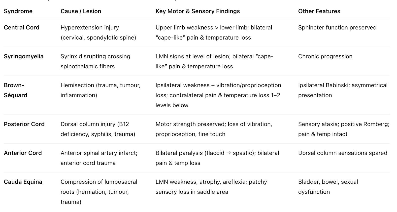
Brown-Séquard syndrome, hemisection of one side of cord side affects;
- Corticospinal tract Ipsilateral weakness
- Dorsal column Ipsilateral ⬇️ proprioception, vibration, fine touch
- Spinothalamic tract Contralateral ⬇️ pain, temperature
Horner's syndrome
△ ptosis, miosis and ipsilateral face anhidrosis due to disturbance to the sympathetic supply to the eye and face. These sympathetic fibres: Start in the hypothalamus ➡ Descend to synapse with preganglionic sympathetic neurons in the lateral horn of T1 to T4 ➡ Fibres travel upward through the sympathetic chain ➡ Some pass through the stellate ganglion ➡ finally, they synapse in the superior cervical ganglion ➡ postganglionic fibres continue to the eye, eyelid, and face.
Cauda equina syndrome
Cauda equina syndrome describes compression of cord below L1/L2 causing bilateral LMN signs of paralysis and reduced reflexes, sensory loss in a saddle distribution, and bladder, bowel and sexual dysfunction. Immediate MRI and surgical decompression usually within 24-48 hours are required.
Exam-style questions ...
📚 Which of the following describes the features of Brown-Séquard syndrome following a spinal cord hemisection?
- Ipsilateral weakness and contralateral loss of temperature distally
- Bilateral weakness and bilateral loss of sensation
- Isolated lower motor neuron weakness
- Sensory level without motor involvement
📚 A woman sustains a gunshot transecting her spinal cord on the left. Which picture fits Brown‑Séquard syndrome?
- Ipsilateral weakness, ipsilateral loss of proprioception & vibration, contralateral loss of pain & temperature
- Contralateral weakness, ipsilateral proprioception loss, contralateral pain & temperature loss
- Ipsilateral weakness, contralateral proprioception & vibration loss, contralateral pain & temperature loss
- Ipsilateral weakness, contralateral proprioception loss, ipsilateral pain & temperature loss
📚 Damage to which of the following causes Horner’s syndrome in a patient with a Pancoast tumour?
- Posterior root of sympathetic
- Lateral root of sympathetic
- Anterior root of sympathetic
- Edinger Westphal nucleus
📚 A patient post‑hyperhidrosis surgery patient presents with ptosis, miosis, and anhidrosis (Horner’s syndrome). Which spinal cord region is affected?
- Anterior horn
- Lateral horn
- Posterior horn
- Dorsal root ganglia
📚 A patient undergoes right thoracoscopic sympathectomy for axillary hyperhidrosis. Diathermy injury may result in:
- Hoarseness
- Horner’s syndrome
- Raised right hemidiaphragm
- Phrenic nerve injury
📚 A patient presents with ptosis, miosis, and anhidrosis, along with numbness over the medial side of the elbow. Which nerve root is most likely affected?
- C5
- T1
- C7
- T2
📚 A patient with a Pancoast tumour presents with wasting of the intrinsic muscles of the hand. Which nerve root is most likely affected?
- C5
- C7
- T1
- L5
📚 A patient with a Pancoast tumour develops ptosis, miosis and anhidrosis. Where is the underlying injury?
- Vagus nerve
- Recurrent laryngeal nerve
- Sympathetic chain at T1
- Phrenic nerve
📚 A patient undergoes treatment for hyperhidrosis. Horner’s syndrome is caused by injury to which ganglion in front of the neck near the 1st rib?
- Gasserian
- Pterygopalatine
- Stellate
- Ciliary
📚 A patient presents with urine retention and features of cauda equina syndrome. Which would not be expected?
- Urine retention
- Knee hyperreflexia
- Overflow incontinence
- Saddle anaesthesia