Spinal Cord Syndromes
👩⚕️ Now we're moving from stroke presentations to classic spinal cord syndromes including: Brown-Séquard syndrome, Horner's syndrome, Cauda Equine syndrome, Anterior Cord syndrome and Cerebellar disease.
Brown-Séquard syndrome
Brown-Séquard syndrome describes spinal cord hemisection:
- Dorsal column જ⁀➴ Ipsilateral 📉 proprioception, vibration, fine touch
- Spinothalamic tract જ⁀➴ Contralateral 📉 pain, temperature
- Corticospinal tract જ⁀➴ Ipsilateral weakness, ((LMN at but UMN below::At the level of the lesion you damage the anterior horn cell and this gives LMN weakness in that myotome only. Below the level of the lesion you damage the corticospinal tract so this gives UMN weakness in all myotomes below the lesion, on the same side.))
Horner's syndrome
Horner's syndrome describes disturbance to the sympathetic supply to the eye and face resulting in:
- Ptosis,
- Miosis and
- Ipsilateral face anhidrosis
👩⚕️ You need to understand and memorise the 3-Neuron Sympathetic Pathway to the head, to identify where it may have gone wrong in Horner's.
The 3-Neuron Sympathetic Pathway
Damage anywhere along this pathway leads to Horner's syndrome.
1️⃣ First-order (central) sympathetic neuron
Hypothalamus ➡ descends through brainstem ➡ synapses in the intermediolateral cell column (IML) at T1–T4.
2️⃣ Second-order (preganglionic) sympathetic neuron
IML (lateral horn) ➡ exits spinal cord ➡ ascends sympathetic chain passing through the ((stellate ganglion:: The stellate ganglion, aka. the cervicothoracic ganglion, lies at the C7-T1 level, near the neck of the first rib.The stellate ganglion lies on the path of the preganglionic sympathetic fibres but is not the ganglion where they synapse. They simply pass through it while ascending to the superior cervical ganglion.)) ➡ superior cervical ganglion.
3️⃣ Third-order (postganglionic) sympathetic neuron
Superior cervical ganglion ➡ follows internal carotid artery ➡ eye, eyelid, and face
Cauda equina syndrome
Cauda equina syndrome describes compression of cord below L1/L2 causing:
- Bilateral LMN signs of paralysis and reduced reflexes,
- Sensory loss in a saddle distribution, and
- Bladder, bowel and sexual dysfunction.
Immediate MRI and surgical decompression within 24-48 hours are required.
Anterior cord syndrome
... describes damage to the anterior ⅔ of the spinal cord, affecting corticospinal and spinothalamic tracts but preserving dorsal column function, resulting in:
- Spinothalamic tract જ⁀➴ Bilateral 📉 pain, temperature
- Corticospinal tract જ⁀➴ Bilateral weakness
- ((LMN weakness at the level::At the level of the lesion you damage the anterior horn cell which causes LMN weakness in that myotome only. Signs include bilateral flaccid paralysis and areflexia.))
- ((UMN weakness below the level::Everything below that level loses UMN input i.e. the brain cannot control them, so this causes UMN weakness in all myotomes below the lesion, signs including spastic weakness, hyperreflexia and Babinski sign.))
- Dorsal column is preserved so vibration and proprioception are spared
Cerebellar lesions
The cerebellum has two major zones: the midline vermis and lateral hemispheres. Injury to either zone results in different symptoms depending on which is affected.
- Vermis lesions lead to:
- Gait/truncal ataxia, with a broad‑based, unsteady gait
- Dysarthria, with slurred scanning explosive speech
- Nystagmus, gaze-evoked, or jerk nystagmus
- Hemisphere lesions lead to:
- Dysdiadochokinesia, impaired rapid alternating movements of the limbs
- Intention tremor, decomposition of movement on goal‑directed tasks
- Limb ataxia
- Hypotonia
- Limb dysmetria, past-pointing
- Poor coordination on finger‑nose testing
Remember the 💥((DANISH::Dysdiadochokinesia, Ataxia, Nystagmus, Intention tremor, Speech, Hypotonia)) mnemonic for cerebellar signs.
Syringomyelia
… describes a fluid-filled cavity (syrinx) within the central spinal cord, typically in the cervical region, which expands outward and compresses crossing spinothalamic fibres first. As it enlarges, it may involve anterior horn cells and, later, corticospinal tracts. This results in:
- Spinothalamic tract (crossing fibres at the level) જ⁀➴ Bilateral 📉 pain & temperature in a ((“cape-like” distribution::Cape-like distribution describes pain & temperature loss across the shoulders, arms, upper torso, due to disruption of the anterior white commissure in that cervical region.))
If syrinx expands....
- Anterior horn cells જ⁀➴ ((LMN weakness::Muscle wasting, hyporeflexia, fasciculations)) at affected cervical myotome
In late disease...
- Corticospinal tracts જ⁀➴ ((UMN weakness::Spasticity, hyperreflexia, Babinski sign)) in the legs
- Note that dorsal columns (vibration, proprioception) are typically spared
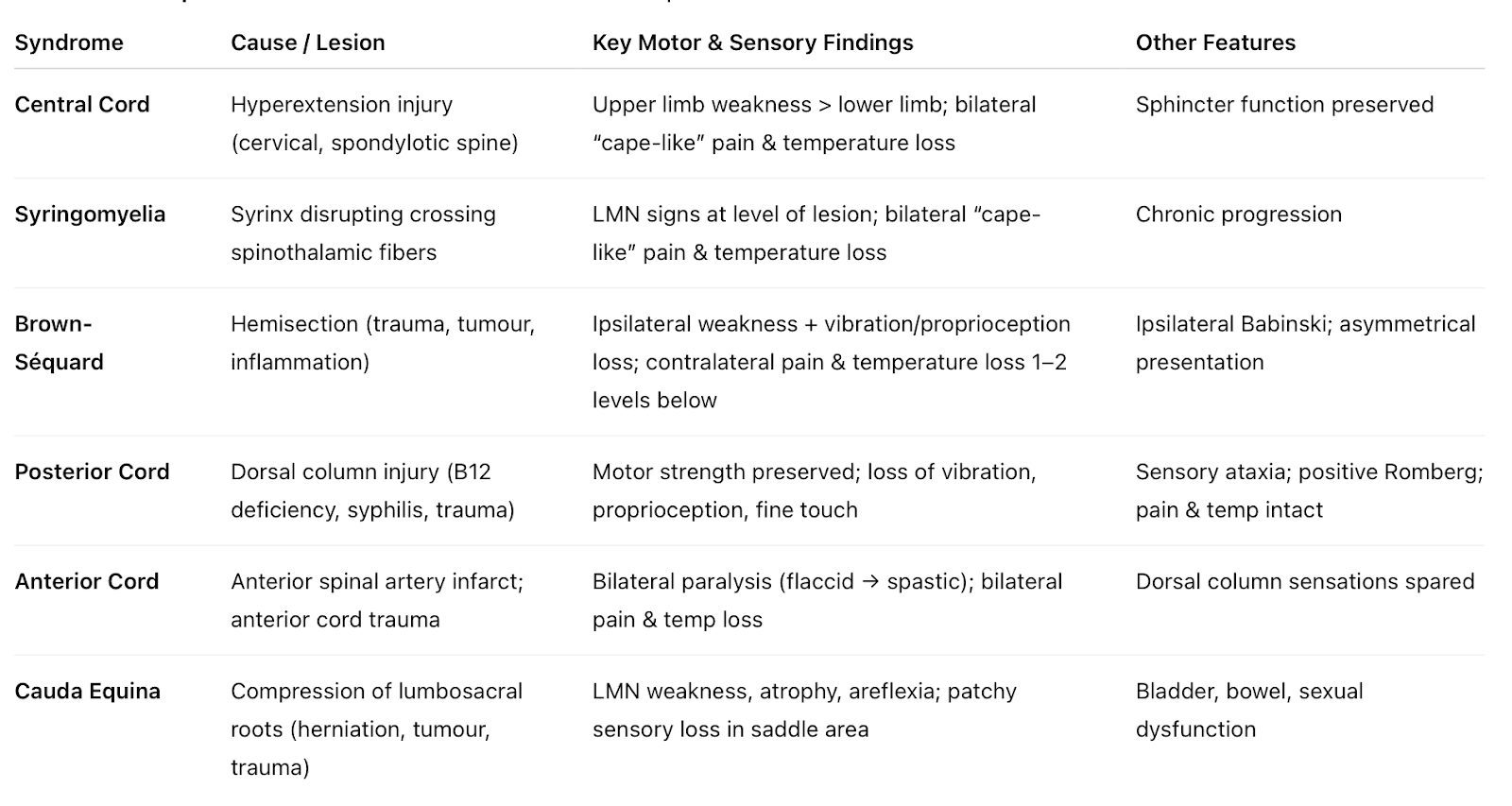
Summary Table of the Spinal Cord Injury Syndromes:
👩⚕️ Yes, you need to know all of these. The Q&As will help consolidate the knowledge!
👩⚕️ Now use these high-yield MCQs to make sure you've got to grips with all these syndromes.
Which of the following describes the features of Brown-Séquard syndrome following a spinal cord hemisection?
- [[Ipsilateral weakness and contralateral loss of temperature distally::Ipsilateral corticospinal tract and dorsal column and contralateral spinothalamic tracts are affected in Brown-Séquard syndrome]]
- ((Bilateral weakness and bilateral loss of sensation::Complete cord transection))
- ((Isolated lower motor neuron weakness::Anterior horn or peripheral nerve lesion))
- ((Sensory level without motor involvement::Posterior cord syndromes))
A woman sustains a gunshot transecting her spinal cord on the left. Loss of which functions fits with Brown‑Séquard syndrome?
- [[Ipsilateral motor, proprioception & vibration, contralateral pain & temperature::Correct]]
- ((Contralateral motor, pain & temperature, ipsilateral proprioception::Incorrect))
- ((Ipsilateral motor, contralateral proprioception, vibration, pain & temperature::Incorrect))
- ((Ipsilateral weakness, pain & temperature, contralateral proprioception::Incorrect))
Damage to which of the following causes Horner’s syndrome in a patient with a Pancoast tumour?
- ((Posterior root of sympathetic::The posterior root carries sensory fibres only and contains no sympathetic outflow, so damage here does not cause Horner’s syndrome.))
- [[Lateral root of sympathetic::Preganglionic sympathetic neurons arise from the intermediolateral cell column (IML) at T1–L2. Their fibres exit as the lateral root, then pass through the stellate ganglion, where a Pancoast tumour can compress them and produce preganglionic Horner’s syndrome.]]
- ((Anterior root of sympathetic::The anterior root contains mostly somatic motor fibres; sympathetic fibres responsible for ocular function leave instead via the white rami communicantes. Damage here does not selectively cause Horner’s syndrome.))
- ((Edinger–Westphal nucleus::This nucleus controls parasympathetic pupillary constriction. Injury here causes a dilated pupil, the opposite of Horner’s syndrome. It does not produce ptosis, miosis, or anhidrosis.))
A patient post‑hyperhidrosis surgery patient presents with ptosis, miosis, and anhidrosis (Horner’s syndrome). Which spinal cord region is affected?
- ((Anterior horn::Contains motor neurons only))
- [[Lateral horn::Contains sympathetic preganglionic neurons running from T1 to L2]]
- ((Posterior horn::Contains sensory relay neurons only))
- ((Dorsal root ganglia::Contains peripheral sensory cell bodies))
A patient undergoes right thoracoscopic sympathectomy for axillary hyperhidrosis. Diathermy injury may result in:
- ((Hoarseness::This would occur with recurrent laryngeal nerve injury, which lies near the tracheoesophageal groove, not at risk during thoracic sympathectomy.))
- [[Horner’s syndrome::Thoracoscopic sympathectomy risks damaging stellate ganglion sympathetic fibres, leading to ptosis, miosis, and anhidrosis on the affected side.]]
- ((Raised right hemidiaphragm::This results from phrenic nerve injury, which lies anterior on the pericardium and is not endangered during posterior sympathetic chain surgery.))
- ((Winged scapula::Occurs due to long thoracic nerve injury, which supplies serratus anterior; not involved in thoracoscopic sympathectomy.))
A patient presents with ptosis, miosis, and anhidrosis, along with numbness over the medial side of the elbow. Which nerve root is most likely affected?
- ((C5::Involved in shoulder abduction))
- [[T1::Involved in sympathetic supply to the eye (Horner’s) and medial arm sensation]]
- ((C7::Supplies triceps and wrist extensors))
- ((T2::Rarely isolated))
A patient with a Pancoast tumour presents with wasting of the intrinsic muscles of the hand. Which nerve root is most likely affected?
- ((C5::Supplies deltoid/rotator cuff))
- ((C7::Supplies triceps and wrist flexors))
- [[T1::Compression of brachial plexus roots (C8, T1) which supplies intrinsic hand muscles]]
- ((L5::Supplies ankle dorsiflexors))
A patient with a Pancoast tumour develops ptosis, miosis and anhidrosis. Where is the underlying injury?
- ((Vagus nerve::Parasympathetic innervation, not sympathetic chain))
- ((Recurrent laryngeal nerve::Causes hoarseness, not Horner’s))
- [[Sympathetic chain at T1::Compression of stellate ganglion’s sympathetic chain at T1]]
- ((Phrenic nerve::Causes diaphragmatic paralysis, not Horner’s))
A patient undergoes treatment for hyperhidrosis. Horner’s syndrome is caused by injury to which ganglion in front of the neck near the 1st rib?
- ((Gasserian::Trigeminal sensory ganglion))
- ((Pterygopalatine::Synapse of parasympathetic greater petrosal nerve (from facial CN VII) to lacrimal gland))
- [[Stellate::Sympathetic ganglion of C7 to T1, near 1st rib, to the head, neck, heart and upper limbs]]
- ((Ciliary::Includes synapse of parasympathetic oculomotor CN III to short ciliary muscles))
The stellate ganglion is formed by the fusion of which sympathetic ganglia?
- ((C5/C6::Incorrect))
- ((C6/C7::Incorrect))
- [[C7/T1:: The stellate ganglion (aka. the cervicothoracic ganglion) lies at the C7-T1 level, near the neck of the first rib]]
- ((T1/T2::Incorrect))
The stellate ganglion (also called the cervicothoracic ganglion) lies at the C7-T1 level, near the neck of the first rib. It is formed by the fusion of the inferior cervical ganglion at C7/C8 and the first thoracic sympathetic ganglion T1. It provides sympathetic innervation to the head, neck, heart, and upper limbs. Stellate ganglion blocks are used for conditions like complex regional pain syndrome and vasospastic disorders.
A patient presents with urine retention and features of cauda equina syndrome. Which would not be expected?
- ((Urine retention::Cauda equina syndrome affects the S2–S4 nerve roots, disrupting parasympathetic fibres to the detrusor and somatic fibres to the external urethral sphincter, leading to a flaccid areflexic bladder. This is expected.))
- [[Knee hyperreflexia::Cauda equina syndrome produces LMN signs, so reflexes are reduced or absent. Hyperreflexia would not be expected.]]
- ((Overflow incontinence::Overflow incontinence occurs because the bladder becomes atonic and overfills, causing passive leakage. This is expected in CES.))
- ((Saddle anaesthesia::Loss of sensation in the S2–S4 dermatomes is an expected feature.))
Cauda equina syndrome describes compression of cord below L1/L2 causing:
- Bilateral LMN signs of paralysis and reduced reflexes
- Sensory loss in a saddle distribution, and
- Bladder, bowel and sexual dysfunction.
Immediate MRI and surgical decompression usually within 24-48 hours are required.
A man has back pain and lower‑limb weakness due to a central disc prolapse causing LMN signs. Diagnosis?
- [[Anterior cord syndrome::A central disc prolapse compresses the anterior spinal cord, damaging the corticospinal tracts (→ LMN flaccid weakness at the level) and spinothalamic tracts (→ loss of pain & temperature below the lesion). Proprioception is preserved.]]
- ((Central cord syndrome::Causes greater upper-limb weakness than lower limbs with cape-like pain/temperature loss. Not consistent with isolated lower-limb LMN signs.))
- ((Brown-Séquard syndrome::Gives ipsilateral UMN weakness, proprioceptive loss, and contralateral pain/temperature loss—a hemisection pattern, not from a central disc prolapse.))
- ((Posterior cord syndrome::Causes loss of proprioception and ataxia, not LMN weakness or pain/temperature loss.))
A woman presents with an unsteady gait but no dysmetria or past‑pointing. Most likely site of lesion?
- ((Upper motor neuron::UMN lesions cause weakness, spasticity, and hyperreflexia, not isolated gait ataxia.))
- ((Posterior horn of spinal cord::Posterior horn lesions cause sensory loss and pain/temperature deficits, not pure gait ataxia.))
- [[Cerebellum::Gait ataxia without limb dysmetria or past-pointing is classic for a vermis (midline) cerebellar lesion, which controls stance and gait.]]
- ((Basal ganglia::Basal ganglia disease causes bradykinesia, rigidity, or tremor i.e. Parkinsonism))
The cerebellum has two major zones: the midline vermis and lateral hemispheres. Injury to either zone results in different symptoms depending on which is affected.
- Lesions in the vermis lead to gait/truncal ataxia, dysarthria and nystagmus
- Lesions in the hemispheres lead to dysdiadochokinesia, intention tremor, limb hypotonia, limb dysmetria (past-pointing) and poor coordination on finger‑nose testing.
A man has spastic paralysis and an extensor plantar reflex. Where is the lesion?
- ((Posterior (dorsal) horn of spinal cord::The dorsal horn is the principal site for termination of primary afferent sensory fibers from the dorsal root ganglia, mediating the initial synaptic processing of somatic sensory modalities including nociception, temperature, and fine touch.))
- ((Cerebellum::💥DANISH: Dysdiadochokinesia, Ataxia, Nystagmus, Intention tremor, Scanning slurred speech, Hypotonia))
- ((Basal ganglia::Consists of corpus striatum (caudate nucleus, putamen, globus pallidus), claustrum, amygdaloid nucleus and thalamus::lesion here causes signs of Parkinsonism including cog‑wheel rigidity, resting tremor, bradykinesia))
- ((Anterior horn of spinal cord::Ventral (anterior) horn of grey matter contains the cell bodies of lower motor neurons which send axons out through the ventral roots of spinal nerves to innervate skeletal (voluntary) muscles; lesion here causes LMN signs such as flaccid paralysis, fasciculations, atrophy))
- [[None of the above::Spasticity, hypertonia, hyperreflexia, and extensor plantar all indicate UMN damage]]
Which structure is primarily responsible for autonomic nervous system control?
- ((Thalamus::Sensory relay))
- ((Medulla::Houses vital reflex centres but under hypothalamic control))
- ((Globus pallidus::Part of basal ganglia, involved in motor control))
- [[Hypothalamus::Regulator of autonomic, endocrine, and homeostatic function]]
Which function is lost in the event of injury to the dorsal root ganglion?
- ((Voluntary motor control::Motor control is mediated by ventral horn motor neurons and the ventral roots, not the dorsal root ganglia.))
- ((Autonomic regulation::Although autonomic fibres pass through spinal nerves, the cell bodies of autonomic neurons are located in the lateral horn (preganglionic) and autonomic ganglia (postganglionic), not in the dorsal root ganglia.))
- [[Sensory perception::Dorsal root ganglia contain the cell bodies of first-order sensory neurons, transmitting pain, temperature, touch, and proprioception.]]
- ((Reflex motor output::The afferent limb of a reflex arc may be affected by DRG injury, but motor outputitself originates from the ventral horn and is not lost directly.))
An elderly man develops cog‑wheel rigidity and bradykinesia. Which structure is most likely affected?
- ((Posterior horn of spinal cord::Posterior horn lesions cause sensory loss and pain/temperature deficits))
- ((Cerebellum::Would cause DANISH signs))
- [[Basal ganglia::Bradykinesia means slowness in starting and performing movements. Cogwheel rigidity feels like a ratchet-like catch when you passively move a patient’s limb. Along with a resting tremor, these are the classic signs of Parkinsonism.]]
- ((Anterior horn of spinal cord::Causes LMN weakness and muscle wasting, hyporeflexia, fasciculations))
The basal ganglia (striatum, globus pallidus, substantia nigra, and subthalamic nucleus) control movement through two systems:
- The direct pathway helps start and smoothen movements.
- The indirect pathway slows or stops movements.
Normally, dopamine from the substantia nigra boosts the direct pathway and inhibits the indirect pathway, allowing smooth motion. In Parkinson’s disease, dopamine-producing neurons die, so movement starts slowly (bradykinesia) and muscles become stiff (rigidity).
How does transcutaneous electrical nerve stimulation (TENS) relieve chronic pain?
- [[Inhibition of posterior horn of grey matter::TENS activates Aβ sensory fibres (touch, vibration) which inhibits transmission from pain-carrying C and Aδ fibres.]]
- ((Inhibition of reticular activating system::RAS controls wakefulness, alert, sleep-wake cycle, suppress noise))
- ((Inhibition of Sodium channel activity::Local anaesthetics block voltage-gated Sodium channels in peripheral axons))
- ((Release of Substance P::Substance P inhibitors are being investigated for use as analgesics))
TENS is based on the "Gate Control Theory" of pain: TENS stimulates Aβ touch/pressure fibres, which inhibits transmission of pain via the Aδ and C fibres so pain feels less intense. This occurs via interneurons in the posterior horn (substantia gelatinosa).
- Aβ fibres: Transmission of touch and pressure
- Aδ fibres: Transmission of acute, localised pain, and temperature
- Aγ fibres:Transmission of motor proprioception information
- B fibres: Autonomic fibres
- C fibres: Slow transmission of mechanothermal stimuli
An elderly man with an Arnold-Chiari malformation develops syringomyelia. Which of the following fibre tracts is most likely disrupted?
- [[Lesion in spinothalamic tract::In Arnold-Chiari malformation, the central cord cavitation interrupts decussating pain/temperature fibres]]
- ((Meningocele::Meningocele contains CSF sac only, no neural tissue involvement))
- ((Meningomyelocele::Meningomyelocele contains protrusion of cord elements, not specific to syrinx formation))
- ((Hydrocephalus::Hydrocephalus may accompany Chiari but not the direct cause of syrinx))
- ((Syringobulbia::Syringobulbia describes extension into brainstem, not the initial spinal cord lesion))
Syringomyelia produces a “cape‑like” loss of pain and temperature as syrinx expansion in the central cord interrupts the crossing spinothalamic fibres in the anterior commissure.
A 2‑year‑old child with Arnold-Chiari malformation develops syringomyelia. Which spinal tract is first affected?
- [[Spinothalamic tract::Crossing pain and temperature fibres decussate in the central canal region, which is the first area expanded by a syrinx, so these fibres are damaged first.]]
- ((Corticospinal tract::Motor fibres are located more laterally and are affected only in late or advanced syringomyelia.))
- ((Dorsal column::The dorsal column carries vibration and proprioception, located posteriorly, so usually spared until late disease.))
- ((Spinocerebellar tract::Spinocerebellar tract fibres lie peripherally in the cord and are not affected early in syringomyelia.))
- ((Sympathetic pathways::Sympathetic fibres are located in the lateral horn, not central, so typically affected much later.))